With our Telehealth services, DISC is ready to take care of you. Learn More »
Understanding the Parts of Your Spine

Understanding the Parts of Your Spine

By Mark Wang, M.D.

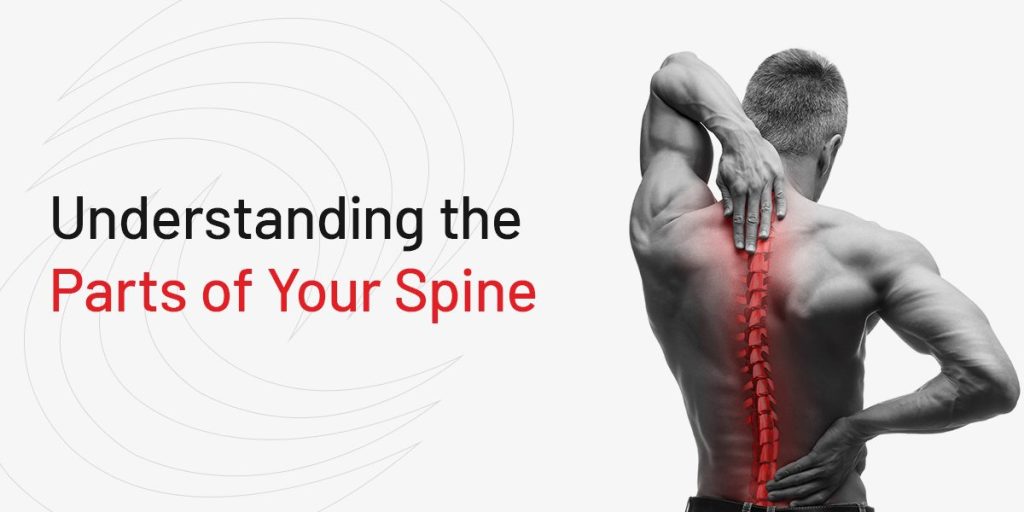
The spine is an important part of the body that provides support and allows for proper mobility. The spinal column is the supportive core of our bodies, while the spinal cord bridges the brain with the rest of the body. Many people may only notice or pay attention to the spine unless there is an issue or pain. Understanding the spinal column and spinal cord anatomy can help you protect yourself from potential injuries. Additionally, understanding the parts of the spine can also help you understand where pain or discomfort may be originating from.

Visit Here to Schedule An Appointment

How the Spine Works: Three Main Functions

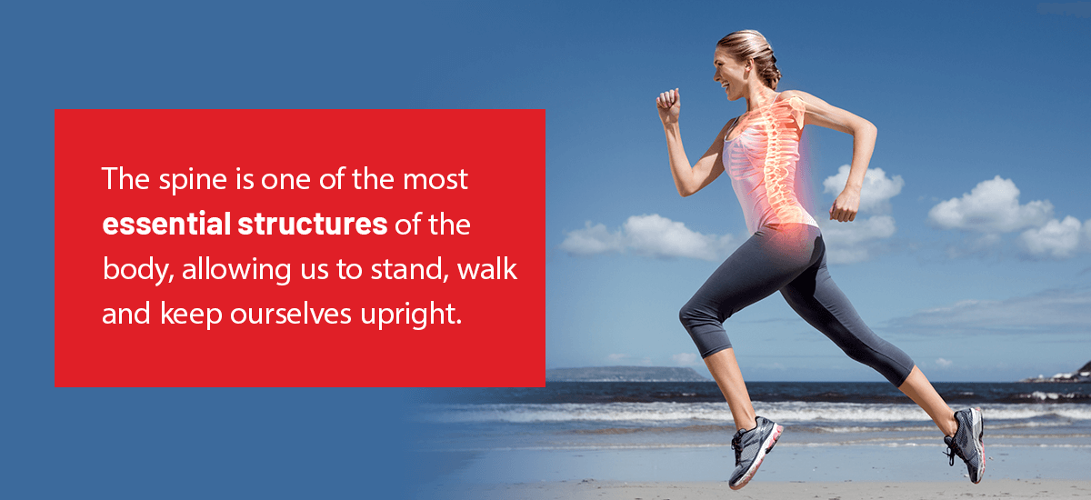
The spine is one of the most essential structures of the body, allowing us to stand, walk and keep ourselves upright. First and foremost, the spine provides your body with proper support. The support of the spine is essential to various bodily movements and functions, such as standing and walking.Additionally, your spine allows for flexible movements, such as bending and twisting. Another function of the spine is that it protects the delicate spinal cord from potential damage. The spinal cord is a collection of nerves that connect the brain to the rest of the body, allowing for various movements and processes. The spinal cord helps the body and internal organs function properly. A healthy spine is a key aspect of leading a healthy lifestyle.

Woman Running While We Can See X-Ray of Spinal Structure

The spinal cord helps the body to move, determine the position of the legs and arms and even feel various stimuli, including hot, cold, sharp or dull sensations. Additionally, the spinal cord plays a vital role in controlling bodily functions like breathing and using the bathroom. Blood pressure, body temperature and heart rate are also regulated and controlled by the spinal cord and brain.

Spinal Anatomy

The spine is a complex structure that is responsible for providing support to the body. There are numerous areas of the spine that each has a unique function and helps the body perform vital processes. The various regions of the spinal column and cord work together to send messages from the brain to different areas of the body.

Spinal Column Regions

The back has four main parts, including the sacral, lumbar, thoracic and cervical regions. These parts of the spine make up the back anatomy, and each serves a unique purpose.

The sacrum is composed of five bones fused together into a triangular shape and situated behind the pelvis. The sacrum fits between the two hip bones and connects the pelvis to the spine. Immediately below the sacrum is the coccyx, also known as the tailbone, consisting of five fused bones.

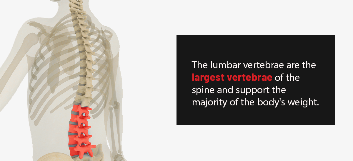
The next portion of the spine is the lumbar spine, which consists of five vertebrae. These vertebrae are the largest of the spine and are responsible for carrying and supporting the majority of the body’s weight. The lumbar spine allows for a wider range of mobility than the thoracic spine but less mobility than the cervical spine.

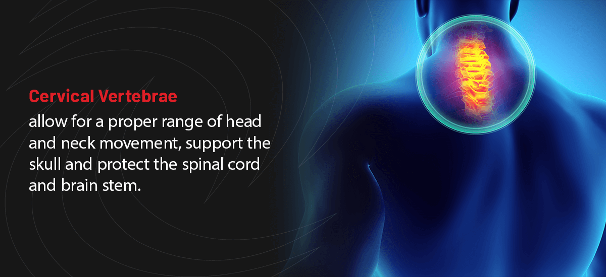
The 12 thoracic vertebrae are located above the lumbar spine and below the last cervical vertebra. The thoracic vertebrae have longer spinous processes and are larger than the cervical bones. The uppermost portion of the spine makes up the neck region and is known as the cervical spine. The cervical spine comprises seven vertebrae that help protect the upper spinal cord and the brain stem.

Parts of the Spinal Cord

Parts of the Spinal Cord

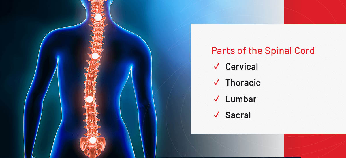
The spinal cord is an important structure between the brain and the body that carries nerve impulses between the brain and the spinal nerves. Similar to the spinal column, the spinal cord is divided into four sections, including the sacral, lumbar, thoracic and cervical regions.

Spinal Curves

The spine shows unique curves when viewed from the side. These curves are known as either kyphotic or lordotic curves. A kyphotic curve is a convex spinal curve found in the sacral or thoracic spinal segments. On the other hand, a lordotic curve is a concave spinal curve found in the lumbar or cervical portions of the spine.

How Many Vertebrae Are There?

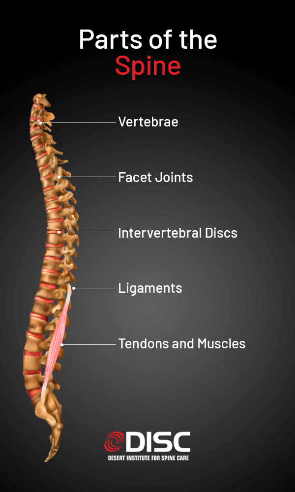
The spinal column is made of 33 vertebrae, which are individual bones that interlock and connect with one another. Vertebral anatomy is divided and numbered depending on the region of the spine, including the vertebrae of the coccygeal, sacral, lumbar, thoracic and cervical regions. The top 24 vertebrae are mobile, while the vertebrae of the coccyx and the sacrum are fused. The spinal column is a tubular or tunnel structure protecting the spinal cord and sensitive spinal nerves from injury.

  • Cervical Vertebrae

    The cervical spine is the neck region that consists of seven vertebrae labeled C1 through C7 from top to bottom. The cervical vertebrae allow for a proper range of head and neck movement, support the skull and protect the spinal cord and brain stem. C1 is the first cervical vertebra, known as the atlas. C1 is a ring-shaped structure that helps support the skull.

    The C2 is known as the axis, which is circular and features a blunt and tooth-like structure that projects upward into the C1. C1 and C2 allow the head to turn and rotate. The remaining cervical vertebrae are C3 through C7 and are box-shaped with small spinous projections extending from the back.

    Cervical Vertebrae
  • Thoracic Vertebrae

    There are 12 thoracic vertebrae that lie under the last cervical vertebra and are labeled T1 through T12 from top to bottom. T1 is the smallest of the thoracic vertebrae, while T12 is the largest. Compared to the cervical vertebrae, the thoracic vertebrae are larger and have longer spinous processes. The thoracic vertebrae are limited in mobility as they are securely attached to the sternum and ribs.

  • Lumbar Vertebrae

    The lumbar spine is comprised of five vertebrae that are abbreviated L1 through L5. The lumbar vertebrae are the largest vertebrae of the spine and support the majority of the body's weight. The lumbar spine allows for greater mobility than the thoracic spine but less mobility than the cervical spine. The lumbar facet joints provide significant mobility and extension but limit rotation.

    The facet joints contain cartilage that allows the vertebrae to slide along one another without pain or damage. Additionally, the facet joints also let you rotate, twist and turn, providing flexibility and stability. Because cartilage wears down over time, the facet joints may be susceptible to arthritis, causing chronic discomfort or pain in the back or neck.

    Lumbar Vertebrae
  • Sacral Vertebrae

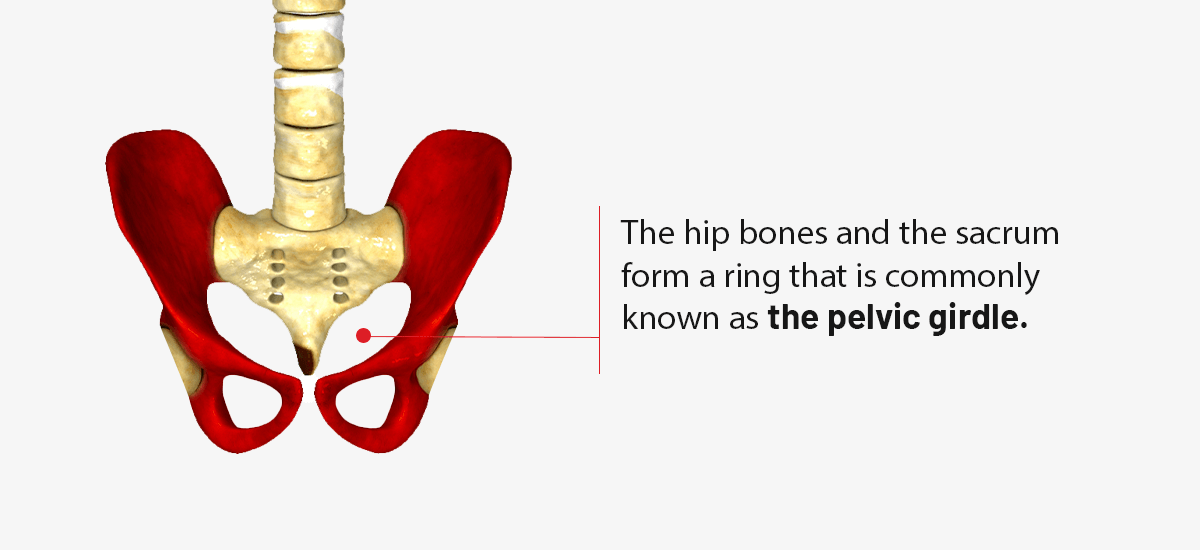
    The sacrum is situated directly behind the pelvis and is composed of five bones labeled S1 through S5. The sacral bones are fused together and form a triangular shape. The sacrum connects the spine to the pelvis and fits between each hip bone. The final lumbar vertebra is known as L5 and moves in conjunction with the sacrum. Because the sacral vertebrae are fused, they do not allow for individual mobility. The hip bones and the sacrum form a ring that is commonly known as the pelvic girdle.

    Sacral Vertebrae
  • Coccygeal Vertebrae

    The coccyx, which is more commonly referred to as the tailbone, is comprised of four vertebrae fused together. The ligaments and pelvic floor muscles attach to the tailbone. Additionally, several ligaments, muscles and tendons connect to the tailbone. While the tailbone is much smaller than the sacrum, it plays an essential role in bearing weight. The tailbone is designed to support your body's weight while you are sitting.

    Coccygeal Vertebrae

Anatomy and Function of the Spinal Cord

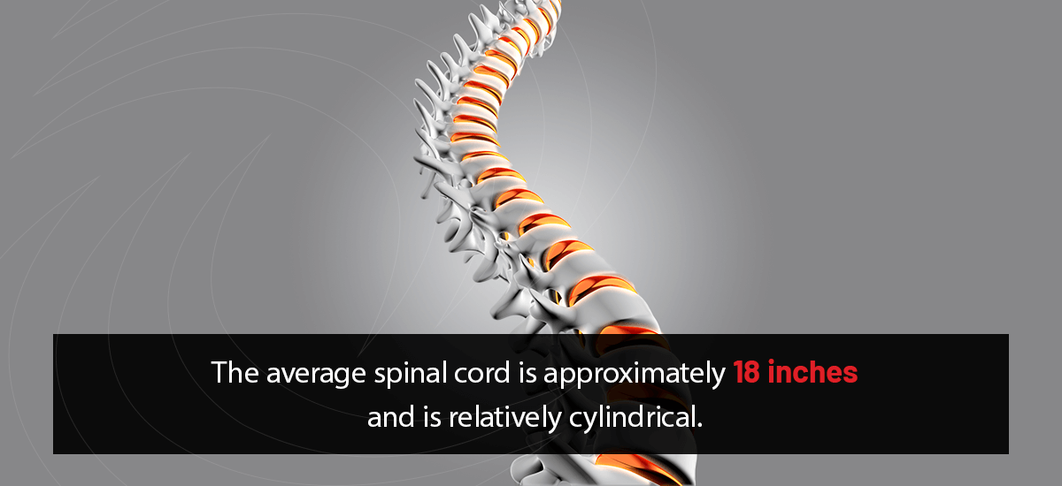
The spinal cord runs from the highest neck bone to the highest vertebra of the lower back, or from C1 to L1. The average spinal cord is approximately 18 inches and is relatively cylindrical. The brain and spinal cord are protected by three layers of membranes known as the meninges. The pia mater is the delicate inner layer, the arachnoid is the middle layer and the dura mater is the tougher outer layer.

The Average Spinal Cord is Approximately 18 Inches
The joints, skin and internal organs send specialized neurons to the spinal cord that help the brain recognize various sensations, including touch, pain, temperatures and vibrations. These messages are conveyed to the brain from the spinal cord through the lemniscal pathway or spinothalamic tract. Another important component of the spinal cord are the rexed lamina, including the:

  • Rexed lamina I: Lamina I is comprised of a thin layer of cells that cap the uppermost portion of the dorsal horn with an array of nonmyelinated axons and small dendrites. The cells in lamina I respond to thermal and noxious stimuli.
  • Rexed lamina II: Lamina II is composed of tightly packed interneurons and also responds to noxious stimuli. Many of the neurons in this lamina receive various information from the sensory dorsal root ganglion cells and the descending dorsolateral fasciculus.
  • Rexed lamina III: Lamina III is involved in perceiving the sensation of light, touch and proprioception — the body’s perception or awareness of its movement and position. The cells within this layer connect with the cells located in IV, V and VI.
  • Rexed lamina IV: This lamina is involved in relaying and processing non-noxious sensory information. These cells connect with the cells located in lamina II and partially correspond to the nucleus proprius.
  • Rexed lamina V: Lamina V relays sensory information, including potentially harmful sensations. This information is sent to the brain through the spinothalamic and contralateral tracts. This lamina also receives descending information from the brain through the rubrospinal and corticospinal tracts.
  • Rexed lamina VI: This lamina contains numerous small interneurons that are involved in promoting spinal reflexes. Lamina VI also receives important sensory information from the muscle spindles. Information is sent to the brain using the ipsilateral spinocerebellar pathways.
  • Rexed lamina VII: A large heterogeneous zone that varies throughout the length of the spinal cord that receives information from lamina II, lamina VI and viscera. This lamina gives rise to cells that are directly involved in the autonomic system.
  • Rexed lamina VIII: Lamina VIII varies depending on the spinal cord level, but it is most prominent in cervical and lumbar enlargements. These cells are involved in modulating motor output to the skeletal muscle.
  • Rexed lamina IX: The size and shape of lamina IX vary between the various levels of the spinal cord. There are also distinct groups of motor neurons that supply the skeletal muscle with nerves.
  • Rexed lamina X: Lamina X surrounds the central canal. The axons cross over from one side of the spinal cord to the other side.
Diagram of the Parts of the Spine

Parts of the Spine and Their Function

The various parts of the spine provide support and enable unique functions and mobilities. With time, the various parts of the back may wear down or degenerate. The degradation of healthy joints and aspects of the spine can lead to limited mobility, inflexibility and even acute or chronic back pain. In addition to chronic back pain, some of the most common spinal conditions include degenerative disc diseaseherniated discsfacet joint syndromedisc tears and more.

  • 1. Vertebrae

    The vertebrae are important spinal bones that consist of a cylindrical body in the front and a bony ring in the back. The main body of the vertebra is a stacking agent that helps to create the entire spinal column. The vertebrae help provide basic weight support to the spine. The delicate spinal cord also runs through the tunnel formed by the stacked vertebrae. The vertebral anatomy includes:

    • Pedicles: Each vertebra features two cylinder-shaped projections of bone known as pedicles. These pedicles of hard bone protrude outwards from the back part of the vertebral body and help protect the sides of the spinal nerves and spinal cord. The pedicles of the spine also act as a bridge that joins the back and front parts of the vertebra.
    • Laminae: The vertebral laminae are bony sheets or ridges that help connect various morphological landmarks of the vertebrae, including zygapophyses and diapophyses. The laminae typically feature some variation throughout the spinal column.
    • Transverse processes: A transverse process is a small bony projection off the left and right sides of each vertebra. There are two transverse processes of each vertebra that serve as the attachment site for ligaments and muscles of the spine and are the point of articulation of the ribs.
    • Spinous processes: A spinous process is a bony projection that is on the back of each vertebra. The spinous process begins to protrude from where the laminae of the vertebral arch join and acts as a point of attachment for various spinal ligaments and muscles.
    • Endplates: The endplates of the vertebral body are anatomically discreet structures that form the interface between the intervertebral discs and vertebral bodies. The endplates are constituted peripherally by the epiphyseal bone ring and centrally by a layer of cartilage.
    • Intervertebral foramina: The intervertebral foramina are often referred to as the neural foramina and are located between two spinal vertebrae. The lumbar, thoracic and cervical spine all have intervertebral foramina. Numerous structures pass through the foramina.
  • 2. Facet Joints

    The facet joints of the spinal column are situated behind the vertebral body and help the spine twist, bend and extend in various directions. While the facet joints enable movement, they also help restrict excessive motion that may result in hyperflexion or hyperextension. Each vertebra of the spinal column has two facet joints. The superior articular facet joint faces upwards and works similar to a hinge with the inferior articular facet joint.

    The facet joints are similar to other joints in the body as they are surrounded by connective tissue, which provides synovial fluid for the joint's nourishment and lubrication. The surfaces of the facet joints are coated in cartilage which protects the joints and allows for smooth movement. Because the facet joints feature interconnected characteristics, they play an essential role in keeping the spinal column stable during movement.

  • 3. Intervertebral Discs

    In between each vertebra, there is a cushion known as the intervertebral disc. The intervertebral disc helps absorb the shock and stress the body may experience during movement while preventing the vertebrae from grinding against one another. The intervertebral discs are also the largest bodily structures that do not have a vascular supply. Each intervertebral disc can also absorb necessary nutrients through the process of osmosis. Each intervertebral disc is comprised of two parts, including the nucleus pulposus and annulus fibrosis.

    • Nucleus pulposus: The nucleus pulposus works in unison with the annulus fibrosis to transmit weight and stress from vertebra to vertebra. The nucleus pulposus consists of proteoglycans, collagen and water. When compared to the annulus fibrosis, the nucleus pulposus contains more water. One of the main functions of the nucleus pulposus is that it helps the vertebral disc withstand torsion and compression.
    • Annulus fibrosus: The annulus fibrosis is a sturdy and almost tire-like structure that encases the nucleus pulposus. The annulus helps enhance the spine's ability to rotate and helps to resist compressive stress. The annulus fibrosus consists of sturdy elastic collagen fibers and water. The fibers are oriented at various angles horizontally. The collagen gains strength from strong bundles of protein that are linked together.
  • 4. Ligaments

    The tendons and ligaments of the spine are bands of fibrous connecting tissue that adhere to the bones. A ligament is designed to connect multiple bones together while stabilizing joints. Ligaments and tendons are relatively elastic and also vary in size. The ligament system in the vertebral column works in conjunction with the muscles and tendons to provide a brace and protect the spine. Ligaments also help to stabilize the joints during movement and rest. The three major ligaments of the spine include:

    • Ligamentum flavum: Meaning "yellow ligament," ligamentum flavum has a yellow coloring due to an abundance of elastin. The elastin pulls the ligament out of the canal when the spine is extended. With age, it is normal for the ligament to lose elastin, which may cause the ligament to encroach on the canal. During lumbar decompression surgery, ligamentum flavum may be removed, as it is often a leading contributor to spinal stenosis.
    • Anterior longitudinal ligament: The anterior longitudinal ligament is a strong band that varies in width and thickness and covers the anterior regions of the vertebral bodies and intervertebral discs through the length of the vertebral column. The deepest fibers are known as the short intersegmental fibers, with the intermediate fibers spanning two or three vertebrae and the superficial fibers spanning three or four vertebrae.
    • Posterior longitudinal ligament: The posterior longitudinal ligament is a strong ligament that forms the anterior wall of the vertebral canal and spans the body from C2 to the sacrum's posterior surface. It features deep intersegmental fibers and more superficial fibers that can span up to four vertebrae.
  • 5. Tendons and Muscles

    Tendons function similarly to ligaments, as these fibrous tissues help secure muscle to bone and are made of collagen fibers. Whether in groups or individually, muscles are supported by fascia, a sturdy, sheath-like connective tissue. The tendon that helps attach the muscle to the bone is part of the fascia.

    The spine's muscular system is complex and has several muscles that play essential roles. The muscles located in the vertebral column help support the spine, provide stability and rotate, flex or extend the spine. Different muscles and muscle groups help to provide mobility to various parts of the body. The sternocleidomastoid muscle in the neck area assists with the mobility of the head. Additionally, the psoas major is a muscle in the lower back area that helps with thigh flexion.

Schedule an Appointment With DISC

The Desert Institue for Spinal Care (DISC) is an orthopedic spine center committed to offering innovative treatments and the highest level of patient care possible. Our team of surgeons and medical experts can help diagnose various spinal conditions and offer spinal treatments and endoscopic spine surgery. We are dedicated to providing compassionate and individualized care for each patient. To learn more about our spinal treatments, contact us online today or call (602) 944-2900.

Schedule an Appointment with DISC for Back and Spine Pain

LEADERS IN MINIMALLY INVASIVE SPINE CARE
1635 E. Myrtle • Suite 400 • Phoenix, AZ 85020
Ph: 602-944-2900 • Fax: 602-944-0064

DISC - Desert Institute for Spine Care